
You never fully appreciate how poor a certain procedure is until a better one comes out and belittles the old one. Most MRI articles spend time telling you what they can do -not what they can’t do… how poor MRIs are is becoming more clear with a new experimental technique called diffusion magnetic resonance imaging.
Foraminal Stenosis
Eur Spine J. 2010 Jul 15. [Epub ahead of print]
Clinical applications of diffusion magnetic resonance imaging of the lumbar foraminal nerve root entrapment.
Eguchi Y, Ohtori S, Yamashita M, Yamauchi K, Suzuki M, Orita S, Kamoda H, Arai G, Ishikawa T, Miyagi M, Ochiai N, Kishida S, Masuda Y, Ochi S, Kikawa T, Takaso M, Aoki Y, Toyone T, Suzuki T, Takahashi K.
abstract here
- They mention: – “Conventional magnetic resonance (MR) imaging (MRI) has been inadequate for evaluating symptomatic foraminal stenosis, because of the high incidence of false-positives found in asymptomatic elderly patients [9]. New diagnostic imaging techniques to detect lumbar nerve root entrapment are urgently required. “
- The article they refer to is:
Aota Y, Niwa T, Yoshikawa K, Fujiwara A, Asada T, Saito T
(2007) Magnetic resonance imaging and magnetic resonance myelography in the presurgical diagnosis of lumbar foraminal stenosis.
Spine 32:896–903 - Foraminal stenosis is common in failed backs but hard to diagnose properly.
Disc Disease Imaging:
Bulging Disc can Hurt and Cause Sciatica/Radiculitis
“There is only a bulging disc falacy” – a study by Jensen found that in chronic sciatica, the big discs disappear after a year but the small mean ones can continue indefinitely with sciatica even without evidence of touching the nerve root:
Spine (Phila Pa 1976). 2006 Jun 15;31(14):1605-12; discussion 1613.
Natural course of disc morphology in patients with sciatica: an MRI study using a standardized qualitative classification system.
Jensen TS, Albert HB, Soerensen JS, Manniche C, Leboeuf-Yde C. abstract here
“Only 3% of bulges [with sciatica] and 38% of focal protrusions improved, whereas 75% to 100% of broad-based protrusions, extrusions, and sequestrations improved (P < 0.0001). ”
Shocking Findings:
- 3/4 of MRIs report bulging discs as normal so you can’t tell.
- Yet 1/2 persistent sciatica MRIs show only a bulge
- Only 10% of bulging disc sciaticas will show nerve root compression.
- Only 10% of acute “bulging discs” sciaticas improve in 14 months
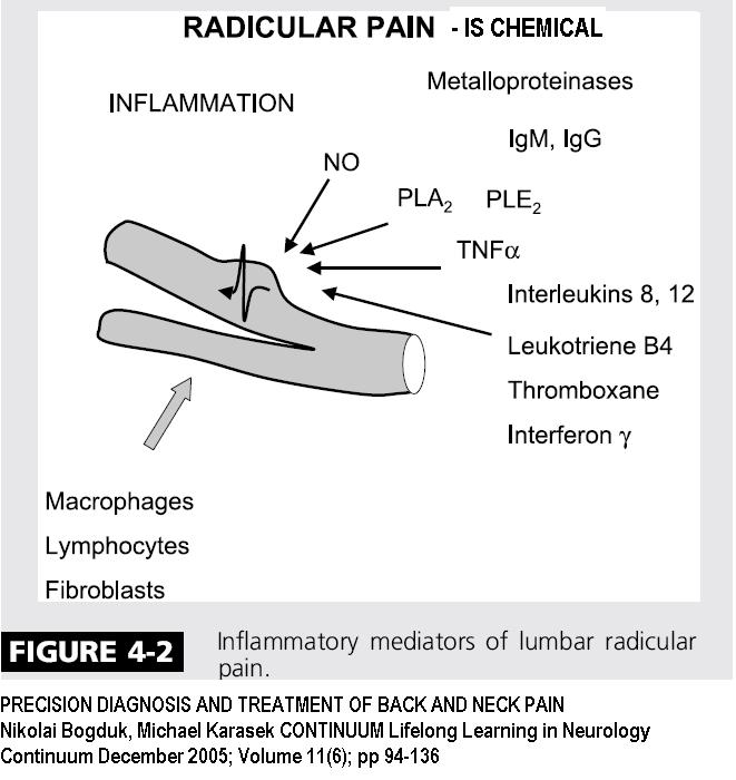
SO HOW USEFUL WAS THE MRI? – NADA – not necessarily because something was not found ,but because a bulge is considered “normal” – but it can’t distinguish between inflamed and not inflamed bulging discs, so their use is poor. It is now realized that much of the pain of sciatica is from irritating chemicals released from damaged discs, NOT the contact, so no actual disc protrusion is even necessary for sciatica – they call that chemical sciatica.
CHEMICAL RADICULITIS not nerve root pressure causes pain
Neck MRIs Often Useless
How useful are neck MRIs?
Eur Spine J (2008) 17:996–1005
Are early MRI findings correlated with long-lasting symptoms following whiplash injury? A prospective trial with 1-year follow-up
Alice Kongsted Æ Joan S. Sorensen Æ Hans Andersen Æ Bjarne Keseler Æ Troels S. Jensen Æ Tom Bendix
free article here
Findings on Neck MRI s (initial and at 3 months post-injury) were not useful in predicting who would have chronic problems.
“In conclusion, MRI is not the answer to a diagnosis in the vast majority of patients developing long-lasting pain after a whiplash injury, and early MRI scans do not predict prognosis.”
Comment – I am sick of radiologists who try to belittle abnormal findings – One I I saw lately:
cervical disc is touching dural sack and spinal cord but [ lying down with head in most comfortable position] not indenting spinal cord – standing with head bent subject was getting pain in legs from bending head forward…
Or saying it’s only a bulging disc where clinical correlation has obviously demonstrated an issue (why would they have an MRI in the first place???)
I’m still waiting for a radiologist to say that the bullet only went one inch into the brain…
Any comments?