Always wondered if imaging sports herniaas much help – apparently not much help in treatment. Older study but their conclusion: – ” MRI revealed a variety of pathological findings in athletes suffering from chronic groin pain, but it was not reliable enough in differentiating between diagnoses requiring conservative or operative treatment.”
J Sports Sci Med. 2007 Mar 1;6(1):71-6.
MRI Findings Do not Correlate with Outcome in Athletes with Chronic Groin Pain.
Daigeler A, Belyaev O, Pennekamp WH, Morrosch S, Köster O, Uhl W, Weyhe D.
http://www.ncbi.nlm.nih.gov/pmc/articles/PMC3778702/pdf/jssm-06-71.pdf
- 19 athletes chronic groin pains
- pain quality was described as
pulling 13
stabbing 5
dull 1 - 7 – accompanying pain in the adductor insertion area (both sides in 2)
- Prior Rx:
2 – hernia repair
1- multiple cortisone injections in the adductor insertion area
1- ultrasound therapy
5- others – physical therapy
MRI findings:
- 7 – bulge on valsalva manouver (bearing down) = tear in the external oblique aponeurosis = posterior abdominal wall insufficiency – 1 later went for surgery; 6 – “achieved complete remission without operative treatment” – not much of a help there then
- 4 – edema in the symphysis (2 had intense pain there) – 1 – edema adductor insertion (though 3 sore there)
- 1 – hydrocele
- 1- osteoma of the left femur,
- 1- enchondroma of the left superior ramus of the pubic bone
- 1- dilated left ureter.
Did mention other studies had seen:
- nerve entrapment (Kopell et al., 1962; Rischbieth, 1986),
- sacroiliac or lumbal abnormalities (Major and Helms, 1997)
- fracture
Their observation: – “It should be noted that 67% (10/15) of the follow-up group were symptom-free after four years simply by reducing or changing their training mode.”
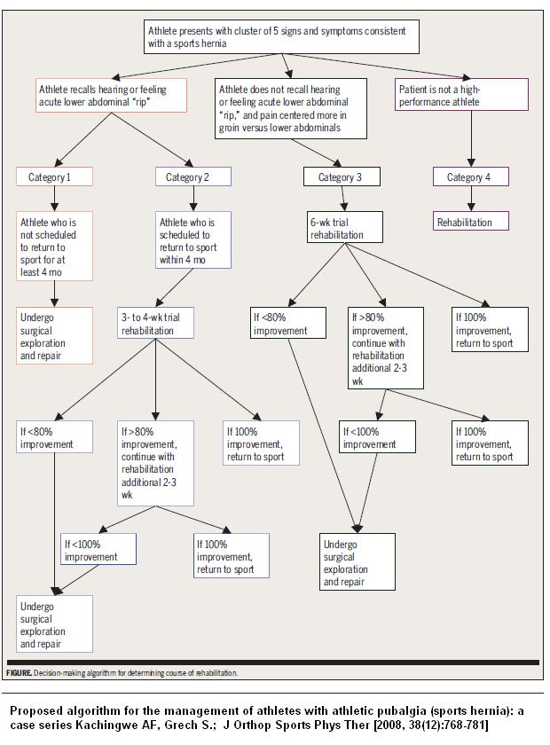
with that in mind, an algorithm was devised in 2008 requiring rehab to fail:

Their conclusion was that
- Physical exam is more sensitive
- MRI did not change treatment plans
“In our opinion most of these findings do not result in a change of the therapeutic algorithm and therefore MRI should be restricted to special indications, when chronic overuse injury of the groin and posterior wall weakness seems to be unlikely as a cause for the groin pain. In almost all cases the localisation of the symptoms corresponded to the sites of pathological findings in MRI (whereas the pain quality was no valid indicator for differentiation e.g. between symphysitis and bulging) further indicating that MRI may not be superior to physical examination in the diagnosis of chronic groin pain.”
Comment – MRI’s again fail to help much with actual treatment but do suggest your clinical assessment might be accurate. Here, the switch has been to doing Ultrasounds – but if MRI’s can’t help you, then I doubt US any better. I suppose it is just reassuring a frank hernia is not missed.

I personally had a sports hernia, and I can agree 100%–while I did get an MRI, it was not used for a full diagnosis. I believe the current diagnosis technique (other than the simple self-test of a pubic probe) is ultrasound while performing the valsalva maneuver. I had a specialist do that and he concluded I did in fact have a sports hernia. Luckily, all better now!