Testing MRI/CT imaged Sacroilitis versus clinical testing and comparing it to positivity of tests with Low back pain came up with low sensitivities and specificities.
Clin Rheumatol. 2008 Oct;27(10):1275-82.
The value of sacroiliac pain provocation tests in early active sacroiliitis. Ozgocmen S, Bozgeyik Z, Kalcik M, Yildirim A.
There were differing results on right and left side so they were displayed separately:
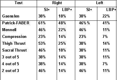
you can see the maximum sensitivity is with the Patrick_faber but only 46-61%.
Looking at the “relative risk” of a test being good:
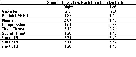
- For specificity – the Patrick-FABER test can be skipped completely as under the needed 2 times RR.
- Tests on the left side seem to be much more specific.
- My favorite, the thigh thrust was OK 2.12(R) – 2.71(L) RR but was seen in only 53% right and 38% left
- The sacral thrust turned out to be the most specific and was as good as combining several tests.
A discussion of SI tests can be found here:
http://www.hwbf.org/hwb/conf/alex47/pex.htm
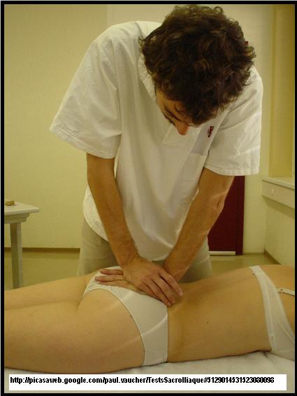
Sacral Thrust test Procedure:
- Patient Prone (on stomach), legs relaxed, semi abducted (leg spread out a bit)
- “[Examiner] stands behind the subject, close to the feet at the lower edge of the table”
- [Examiner] Puts hands over the sacrum applies posterio-anterior pressure to the sacrum
Gaenslen test (pelvic torsion test)
Patient supine (on back), affected side close to the edge and partially off the examination table
examiner stands at the side of the table
“Examiner guides patient flexing the knees to the chest until the subject’s lower back assumes physiologic lordosis. The examiner fixates the contralateral leg and applies a light pressure to the ipsilateral leg causing hyperextension of the hip”
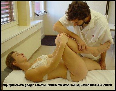
Patrick FABER Test:
Supine
Stands next to the subject
“Subject brings ipsilateral knee into the flexion with lateral malleolus placed over the contralateral knee. Examiner fixates the contralateral ASIS. The examiner applies a light pressure over the ipsilateral knee, gently forcing the knee to contact the table as much as possible
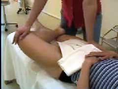
Compression test:
“Side-lying position, affected side is up, close to the side of the table and back
towards the edge of the table. Hips flexed approximately 45°, knees are flexed approximately 90° degrees”
Examiner stands behind the subject
“Examiner’s Folded hands over the anterior edge of the iliac crest and applies
downward pressure”
Can’ find image that is not copy-writed. Can see a netter image at:
http://www.netterimages.com/publication/9781929007875/6-226.htm
Comment – Hope that helps. Looks like you need a high index of suspicion for belt line back pain and consider SI injection if confused.
Any views on the matter?

Hallo. I have a few questions regarding Sacroiliitis. I have had these back pains for the last 25 years (now I’m 49). Doctors in Norway have always told me that it’s nothing wrong except for some herniated disc problems. I have operated for this, but still the same SI Joint pain. Then I lately came over Sacroiliitis. Everything seem to fit with my experience. I only have a few questions to clarify this a litle bit more. 1. My SI Joint pain comes and goes i.e. can come quite sudden, then it become very extreme, at extreme level it last for 1-3 weeks, then I can feel it for 1-2 months, i.e. be carefull etc. Then it can be away for several months, but my experience is that i appears more and more frequent. Does this match with the way Sacroiliitis works? 2. My experience is that when this pain is at the most extreme I can hardly move, the best position for me is with my back on the floar, with my feets on a chear i.e. this position seem to rest/stabilize the SI Joint pain region. To be in a bed or lie on a sofa is very difficult because the stretch on my feet or the slight movements in my back in this position seem to make some slight movements in my SI Joint region and thereby enlarge/prolong these pains. Does this match with the way Sacroiliitis works? Thank you very very much:-)